

China Supplier Dental Surgery Furniture - Planmeca Promax 2D S3 Panoramic X-Ray Unit OPG – JPS DENTAL
China Supplier Dental Surgery Furniture - Planmeca Promax 2D S3 Panoramic X-Ray Unit OPG – JPS DENTAL Detail:
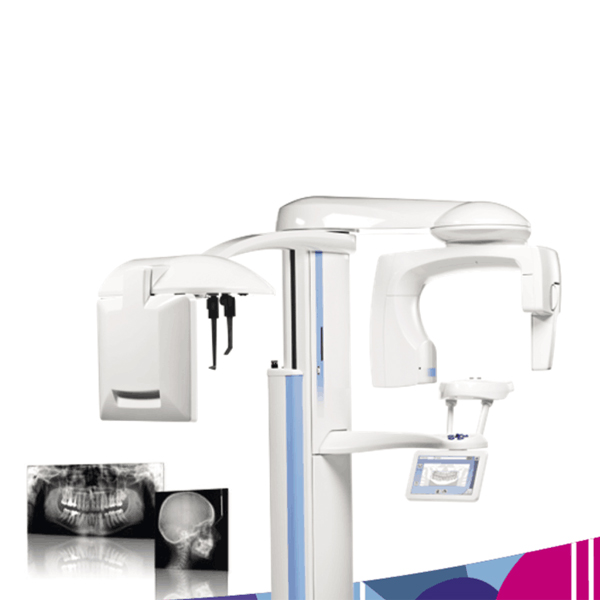
Planmeca ProMax® is a complete maxillofacial imaging system. The design and operation principles are based on the latest scientific research, technological innovations and the most demanding needs of modern-day radiology.
Advanced technology
• Autofocus positions the focal layer automatically for perfect panoramic images
• Dynamic Exposure Control (DEC) measures the patient’s radiation transparency and automatically adjusts exposure values
• Patented SCARA (Selectively Compliant Articulated Robot Arm) technology guarantees an anatomically correct imaging geometry for clear, error-free images
• Easy upgrades – add cephalostat or 3D imaging capability at any time
Effortless use
• Full-view patient positioning with triple-laser patient positioning lights
• Side entry for comfortable access
• Easy-to-use graphical interface
• Versatile Planmeca Romexis® 2D imaging software
• TWAIN support and full DICOM compliance
Planmeca ProMax X-ray unit provides a wide range of extraoral imaging modalities:
- panoramic imaging for dental arch
- maxillary sinus imaging
- temporomandibular joint imaging
- 2D linear tomography
- cephalometry
Open positioning and easy use
- Free view to the patient from all directions
- Three laser positioning laser beams
- Easy access also for wheelchair patients
- Motorized patient positioning and temple supports
- Autofocus feature makes the positioning of the focal layer automatically. Autofocus takes first a
short, low-dose scout image for searching landmarks and calculating the focal layer with the help of a special neural network algorithm. The user can monitor the suggested focal layer adjustment both on the control panel and on the image acquisition preview. The focal layer adjustments can be amended, or the user can simply accept the adjustments and continue to the final exposure.
- Dynamic Exposure Control (DEC) adapts the whole imaging chain individually for each patient’s
physio-anatomical characteristics to produce the optimal contrast and density. Both the X-ray
source and the image receptor are automatically adjusted to produce the optimum image quality.
- Interactive, informative and intuitive colour TFT graphic user interface (GUI)
- Technical factors and selected programs digitally displayed
- Image preview
Optimised image geometry and constant magnification
- Optimised image geometry and constant magnification
- Adjustable form of focal trough
- Automatic compensation for the cervical vertebrae shadow Full digital control
- Re-programmable flash EPROM
- Microprocessor controlled self-diagnostic control system with clear help guiding to correct use and
error messages announcing hardware or software problems
Constant potential, microprocessor controlled resonance mode generator
- Very high operating frequency 80 – 150 kHz
- Maximum ripple 670 Vpp (0.4%, 84 kV)
- Ultra short rise time, < 3 ms
- Very wide exposure parameters range, 1 – 16mA / 54 – 84 kV2(5)
- Low patient dose
- Universal power input including Power Factor Corrector, mains voltage fluctuations automatically
compensated
Reliable mechanical construction
- Small size and light weight, total weight 113 kg (249 lbs)
- Unique 3 joint SCARA (Selectively Compliant Articulated Robot Arm) technology enables
complicated movements and versatile imaging geometries, smooth and quiet micro-step motors
- Telescopic body column without counterweight. Maximum height adjustable.
- Automatic four blade primary collimator
- Available as wall-mounted or free standing
Imaging mode:
- Basic panoramic programs
- Horizontal and Vertical segmenting
- Bitewing panoramic program
- Tomography: Digital tomography, Transtomography
- Child mode in all imaging programs to reduce the dose and to optimise the imaging geometry
Cephalostat
- Planmeca ProCeph “one shot” cephalostat
- Digital Ceph Dimax4 (2 fixed sensors or 1 movable sensor)
DEC (Dynamic Exposure Control):
- Panoramic DEC
- Cephalostat DEC
Autofocus
Additional features:
- Accessory cabinet
Product detail pictures:
Related Product Guide:
The crawler turner adopts the crawler drive design, which can be operated by one person. When it works, the crawler straddles the strip compost pile, and the cutter shaft at the lower end of the frame rotates to mix and turn the raw materials. The operation can be done not only in the open air area, but also in the workshop or greenhouse. China Supplier Dental Surgery Furniture - Planmeca Promax 2D S3 Panoramic X-Ray Unit OPG – JPS DENTAL , The product will supply to all over the world, such as: Cambodia , Ottawa , Riyadh , Cow dung pellet making machine
 By Steven
from Belgium
- 2017.08.16 13:39
By Steven
from Belgium
- 2017.08.16 13:39
Problems can be quickly and effectively resolved, it is worth to be trust and working together.
By Alice
from Turin
- 2017.05.02 11:33
-
100% Original Dental Equipment Manufacturers - ...
-
China OEM Cotton Roll Dispenser - JP-STE-18-D ...
-
Wholesale Dental Apron - Digital Dental X-ray ...
-

Factory Cheap Hot Disposable Dental Equipment -...
-
Lowest Price for Dental Industry - Mobile Stan...
-
Hot Selling for Affordable Dental Furniture - ...