Leave Your Message
Products

Products

Products Categories
Featured Products
Best Price on Dental Supplies Wholesale -
 Plan... Best Price on Dental Supplies Wholesale -
 Plan...
01

Best Price on Dental Supplies Wholesale - Plan...

2016-01-08
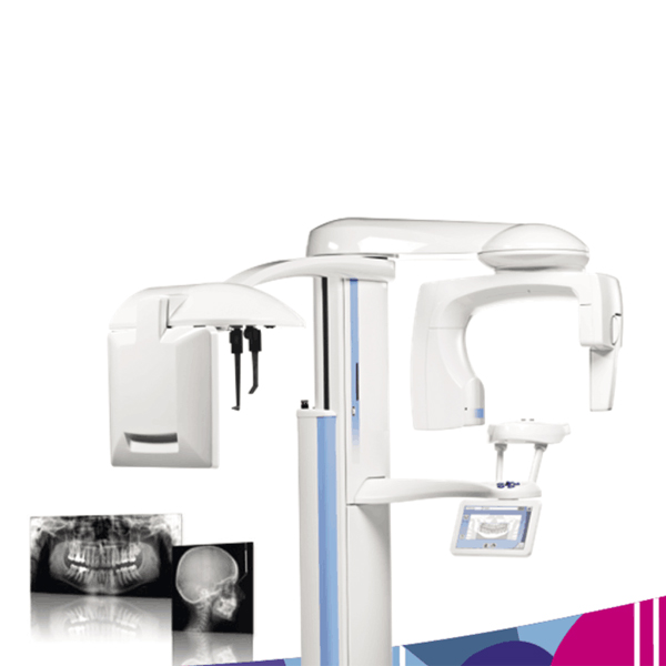
Planmeca ProMax® is a complete maxillofacial imaging system. The design and operation principles are based on the latest scientific research, technological  innovations and the most demanding needs of modern-day radiology. Advanced technology •  Autofocus positions the focal layer automatically for perfect panoramic images •  Dynamic Exposure Control (DEC) measures the patient’s radiation transparency and automatically adjusts exposure values •  Patented SCARA (Selectively Compliant Articulated Robot Arm) technology guarantees an anatomically correct imaging geometry for clear, error-free images •  Easy upgrades – add cephalostat or 3D imaging capability at any time Effortless use •  Full-view patient positioning with triple-laser patient positioning lights •  Side entry for comfortable access •  Easy-to-use graphical interface •  Versatile Planmeca Romexis® 2D imaging software •  TWAIN support and full DICOM compliance Planmeca ProMax X-ray unit provides a wide range of extraoral imaging modalities: panoramic imaging for dental arch maxillary sinus imaging temporomandibular joint imaging 2D linear tomography cephalometry  Open positioning and easy use Free view to the patient from all directions Three laser positioning laser beams Easy access also for wheelchair patients Motorized patient positioning and temple supports Autofocus feature makes the positioning of the focal layer automatically. Autofocus takes first a short, low-dose scout image for searching landmarks and calculating the focal layer with the help of a special neural network algorithm. The user can monitor the suggested focal layer adjustment both on the control panel and on the image acquisition preview. The focal layer adjustments can be amended, or the user can simply accept the adjustments and continue to the final exposure. Dynamic Exposure Control (DEC) adapts the whole imaging chain individually for each patient's physio-anatomical characteristics to produce the optimal contrast and density. Both the X-ray source and the image receptor are automatically adjusted to produce the optimum image quality. Interactive, informative and intuitive colour TFT graphic user interface (GUI) Technical factors and selected programs digitally displayed Image preview Optimised image geometry and constant magnification Optimised image geometry and constant magnification Adjustable form of focal trough Automatic compensation for the cervical vertebrae shadow Full digital control Re-programmable flash EPROM Microprocessor controlled self-diagnostic control system with clear help guiding to correct use and error messages announcing hardware or software problems Constant potential, microprocessor controlled resonance mode generator Very high operating frequency 80 – 150 kHz Maximum ripple 670 Vpp (0.4%, 84 kV) Ultra short rise time, < 3 ms Very wide exposure parameters range, 1 – 16mA / 54 – 84 kV2(5) Low patient dose Universal power input including Power Factor Corrector, mains voltage fluctuations automatically compensated Reliable mechanical construction Small size and light weight, total weight 113 kg (249 lbs) Unique 3 joint SCARA (Selectively Compliant Articulated Robot Arm) technology enables complicated movements and versatile imaging geometries, smooth and quiet micro-step motors Telescopic body column without counterweight. Maximum height adjustable. Automatic four blade primary collimator Available as wall-mounted or free standing  Imaging mode: Basic panoramic programs Horizontal and Vertical segmenting Bitewing panoramic program Tomography: Digital tomography, Transtomography Child mode in all imaging programs to reduce the dose and to optimise the imaging geometry Cephalostat Planmeca ProCeph “one shot” cephalostat Digital Ceph Dimax4 (2 fixed sensors or 1 movable sensor) DEC (Dynamic Exposure Control): Panoramic DEC Cephalostat DEC Autofocus Additional features: Accessory cabinet 
read more
PriceList for Panoramic X-Rays -
 Planmeca Prom... PriceList for Panoramic X-Rays -
 Planmeca Prom...
01

PriceList for Panoramic X-Rays - Planmeca Prom...

2016-01-08
Planmeca ProMax® is a complete maxillofacial imaging system. The design and operation principles are based on the latest scientific research, technological  innovations and the most demanding needs of modern-day radiology. Advanced technology •  Autofocus positions the focal layer automatically for perfect panoramic images •  Dynamic Exposure Control (DEC) measures the patient’s radiation transparency and automatically adjusts exposure values •  Patented SCARA (Selectively Compliant Articulated Robot Arm) technology guarantees an anatomically correct imaging geometry for clear, error-free images •  Easy upgrades – add cephalostat or 3D imaging capability at any time Effortless use •  Full-view patient positioning with triple-laser patient positioning lights •  Side entry for comfortable access •  Easy-to-use graphical interface •  Versatile Planmeca Romexis® 2D imaging software •  TWAIN support and full DICOM compliance Planmeca ProMax X-ray unit provides a wide range of extraoral imaging modalities: panoramic imaging for dental arch maxillary sinus imaging temporomandibular joint imaging 2D linear tomography cephalometry  Open positioning and easy use Free view to the patient from all directions Three laser positioning laser beams Easy access also for wheelchair patients Motorized patient positioning and temple supports Autofocus feature makes the positioning of the focal layer automatically. Autofocus takes first a short, low-dose scout image for searching landmarks and calculating the focal layer with the help of a special neural network algorithm. The user can monitor the suggested focal layer adjustment both on the control panel and on the image acquisition preview. The focal layer adjustments can be amended, or the user can simply accept the adjustments and continue to the final exposure. Dynamic Exposure Control (DEC) adapts the whole imaging chain individually for each patient's physio-anatomical characteristics to produce the optimal contrast and density. Both the X-ray source and the image receptor are automatically adjusted to produce the optimum image quality. Interactive, informative and intuitive colour TFT graphic user interface (GUI) Technical factors and selected programs digitally displayed Image preview Optimised image geometry and constant magnification Optimised image geometry and constant magnification Adjustable form of focal trough Automatic compensation for the cervical vertebrae shadow Full digital control Re-programmable flash EPROM Microprocessor controlled self-diagnostic control system with clear help guiding to correct use and error messages announcing hardware or software problems Constant potential, microprocessor controlled resonance mode generator Very high operating frequency 80 – 150 kHz Maximum ripple 670 Vpp (0.4%, 84 kV) Ultra short rise time, < 3 ms Very wide exposure parameters range, 1 – 16mA / 54 – 84 kV2(5) Low patient dose Universal power input including Power Factor Corrector, mains voltage fluctuations automatically compensated Reliable mechanical construction Small size and light weight, total weight 113 kg (249 lbs) Unique 3 joint SCARA (Selectively Compliant Articulated Robot Arm) technology enables complicated movements and versatile imaging geometries, smooth and quiet micro-step motors Telescopic body column without counterweight. Maximum height adjustable. Automatic four blade primary collimator Available as wall-mounted or free standing  Imaging mode: Basic panoramic programs Horizontal and Vertical segmenting Bitewing panoramic program Tomography: Digital tomography, Transtomography Child mode in all imaging programs to reduce the dose and to optimise the imaging geometry Cephalostat Planmeca ProCeph “one shot” cephalostat Digital Ceph Dimax4 (2 fixed sensors or 1 movable sensor) DEC (Dynamic Exposure Control): Panoramic DEC Cephalostat DEC Autofocus Additional features: Accessory cabinet 
read more
Good quality Cotton Roll -
 Portable X-ray Unit... Good quality Cotton Roll -
 Portable X-ray Unit...
01

Good quality Cotton Roll - Portable X-ray Unit...

2016-01-08
This portable dental X-ray unit is high frequency machine. The body is small, light weight and nearly no radiation. It has excellent image quality, portable storage, save more space. It uses high frequency and DC international power supply. All of the components installed in the central PC board concentrated. Shock, set-up, electron tubes, all of them are insulation vacuum, sealed stereotype protection. There are also manual buttons installed on the surface of shell, as well as batteries and charges. This unit mainly suitable for oral pre-treatment for learning internal organizational structure, root depth and so on, is indispensable in daily life clinic equipment, especially for dental implant surgery. It also can connect with sensor, is great convenience. Checking and Security !  Caution: Please learn relative security measures and functions given in the file or on the unit. * Using the products after checking battery and the location of the device, make sure that is operates properly. * Checking all the power cords whether correctly and connected formally. * Operating the device in the designated order. * Recheck the cone connect with the patient’s face closely. * Keep the device and the cone clean. * Please use or operate the device and parts after taking a necessary periodic inspection. * If you discover some difference in the sensor images even if you inspected its x-ray exposure time in the same way as before, then it means that the radiation of x-ray is abnormal, you must stop using it immediately and allow it to be inspected. * Don’t use or store near the place which is possible to fire. * Don’t use or store where air pressure or temperature or moisture exceeds range. * Keep good ventilation and avoid direct sunlight, protect against the eroding of dust and corrosive air. * Don’t use or store near to chemicals or combustible gas. Tube voltage 60KV Tube current 1.5mA Exposure time 0.1--2.0s Frequency 30KHz Rate power 60VA Distance from skin to cone 130mm Tube focus 0.3mm*0.3mm Battery DC14.8V  6400mAh Charger input voltage AC100V-240V±10% Output voltage DC16.8V Ambient temperature 5℃~40℃ Relative temperature <75%RH Net weight 2.5KG Gross weight 3.5KG Size(mm) 138*165*185 Package size(mm) 365*330*265
read more