Leave Your Message
Products

Products

Products Categories
Featured Products
Manufacturer for Dental Suture -
 Apixia Digita... Manufacturer for Dental Suture -
 Apixia Digita...
01

Manufacturer for Dental Suture - Apixia Digita...

2016-01-08
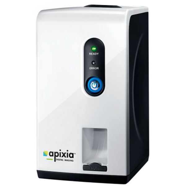
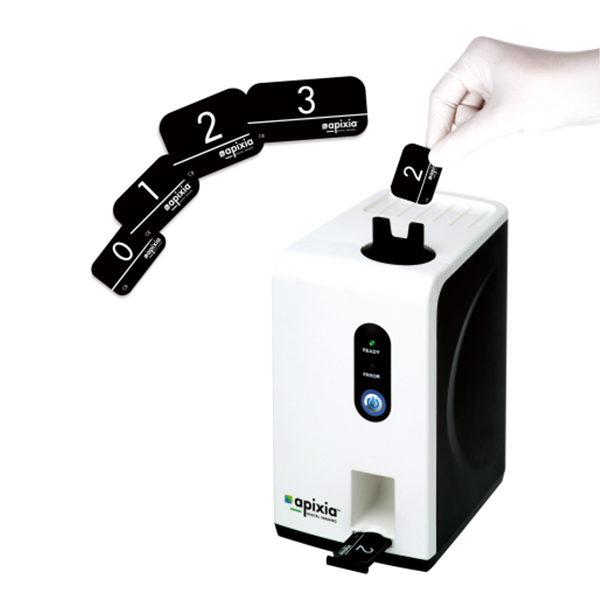
LASER FAST IMAGE PROCESSING Imaging is fast and simple. The smart top-loading design harnesses the power of gravity for simple loading and fuss-free use. In just a short time, the crystal clear radiograph appears on the computer screen, ready for viewing. Simply insert the next exposed plate when ready. FLEXIBLE IN MORE WAYS THAN ONE The Apixia DIGITAL PSP Scanner packs tremendous imaging power into a small package. It is tiny enough to fit into the most crowded dental operatories for handy chair-side use. Imaging with Phosphor plates offers all the flexibility and patient comfort of film, combined with the immediacy and environmental benefits of digital radiography. COMFORTABLE & REUSABLE PHOSPHER PLATES Imaging with Phospher Plates offers all the flexibility and patient comfort of film. Phospher plates are reusable a countless number of times to maximize your investment. 4 optional plate sizes allows for optimum size selection for each patient. SMART AND SIMPLE TOP-LOADING DESIGN Special top-loading design allows for easy image scanning. Just insert and scan any size plate for any type of clinical situation. Quick and easy scanning for easy image viewing on a computer display. 1.Small Tabletop Design 2.High Resolution Scans in a Fraction of the Time 3.Automatic In-line Eraser 4.Powerful, Easy-To-Use Software Included 5.Value-Priced for any Dental Practice Minimum Requirement for Computer Configuration: Windows 7/Windows 8/VISTA/XP (32, 64Bit) 1GB RAM Pentium 4 CPU or much advanced USB 2.0 transmit CD/DVD drive Resolution: >1024*768 40GB for hard drive space High resolution image display HD LCD This machine does not include the computer Digirex Psp Imaging Scanner Specification: Voltage: AC 100-240V, 50/60Hz Scanning time: 10s Resolution: 17 lp/mm Transfer way: USB 2.0 Size of digital X-ray plate: 0# 2# (1# 3# is optional) Built-in cleaning device: Automatic Size: 258*138*183mm Weight: 3.6kg
read more
PriceList for Panoramic X-Rays -
 Planmeca Prom... PriceList for Panoramic X-Rays -
 Planmeca Prom...
01

PriceList for Panoramic X-Rays - Planmeca Prom...

2016-01-08
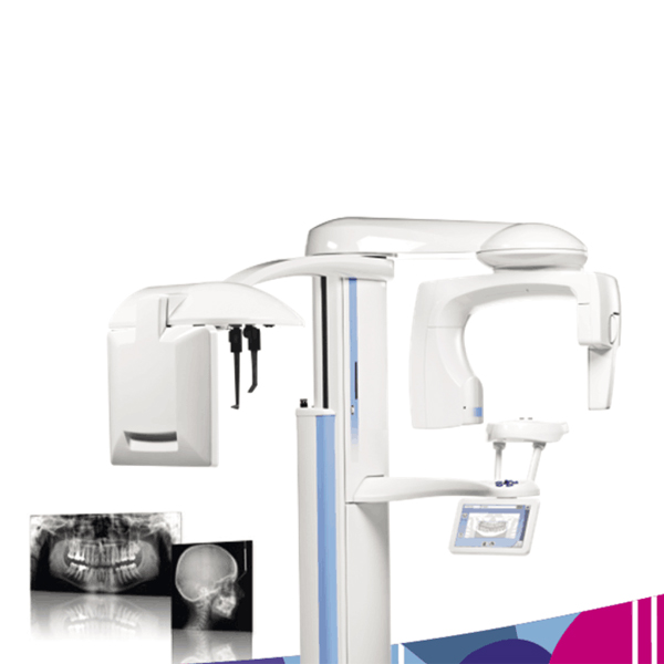
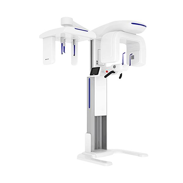
Planmeca ProMax® is a complete maxillofacial imaging system. The design and operation principles are based on the latest scientific research, technological  innovations and the most demanding needs of modern-day radiology. Advanced technology •  Autofocus positions the focal layer automatically for perfect panoramic images •  Dynamic Exposure Control (DEC) measures the patient’s radiation transparency and automatically adjusts exposure values •  Patented SCARA (Selectively Compliant Articulated Robot Arm) technology guarantees an anatomically correct imaging geometry for clear, error-free images •  Easy upgrades – add cephalostat or 3D imaging capability at any time Effortless use •  Full-view patient positioning with triple-laser patient positioning lights •  Side entry for comfortable access •  Easy-to-use graphical interface •  Versatile Planmeca Romexis® 2D imaging software •  TWAIN support and full DICOM compliance Planmeca ProMax X-ray unit provides a wide range of extraoral imaging modalities: panoramic imaging for dental arch maxillary sinus imaging temporomandibular joint imaging 2D linear tomography cephalometry  Open positioning and easy use Free view to the patient from all directions Three laser positioning laser beams Easy access also for wheelchair patients Motorized patient positioning and temple supports Autofocus feature makes the positioning of the focal layer automatically. Autofocus takes first a short, low-dose scout image for searching landmarks and calculating the focal layer with the help of a special neural network algorithm. The user can monitor the suggested focal layer adjustment both on the control panel and on the image acquisition preview. The focal layer adjustments can be amended, or the user can simply accept the adjustments and continue to the final exposure. Dynamic Exposure Control (DEC) adapts the whole imaging chain individually for each patient's physio-anatomical characteristics to produce the optimal contrast and density. Both the X-ray source and the image receptor are automatically adjusted to produce the optimum image quality. Interactive, informative and intuitive colour TFT graphic user interface (GUI) Technical factors and selected programs digitally displayed Image preview Optimised image geometry and constant magnification Optimised image geometry and constant magnification Adjustable form of focal trough Automatic compensation for the cervical vertebrae shadow Full digital control Re-programmable flash EPROM Microprocessor controlled self-diagnostic control system with clear help guiding to correct use and error messages announcing hardware or software problems Constant potential, microprocessor controlled resonance mode generator Very high operating frequency 80 – 150 kHz Maximum ripple 670 Vpp (0.4%, 84 kV) Ultra short rise time, < 3 ms Very wide exposure parameters range, 1 – 16mA / 54 – 84 kV2(5) Low patient dose Universal power input including Power Factor Corrector, mains voltage fluctuations automatically compensated Reliable mechanical construction Small size and light weight, total weight 113 kg (249 lbs) Unique 3 joint SCARA (Selectively Compliant Articulated Robot Arm) technology enables complicated movements and versatile imaging geometries, smooth and quiet micro-step motors Telescopic body column without counterweight. Maximum height adjustable. Automatic four blade primary collimator Available as wall-mounted or free standing  Imaging mode: Basic panoramic programs Horizontal and Vertical segmenting Bitewing panoramic program Tomography: Digital tomography, Transtomography Child mode in all imaging programs to reduce the dose and to optimise the imaging geometry Cephalostat Planmeca ProCeph “one shot” cephalostat Digital Ceph Dimax4 (2 fixed sensors or 1 movable sensor) DEC (Dynamic Exposure Control): Panoramic DEC Cephalostat DEC Autofocus Additional features: Accessory cabinet 
read more
Hot sale Dental Bib Clip -
 Digital 3D OPG Pano... Hot sale Dental Bib Clip -
 Digital 3D OPG Pano...
01

Hot sale Dental Bib Clip - Digital 3D OPG Pano...

2016-01-08
Pano: •Provide you with high diagnosis accuracy and better treatment on diseasesof the full dentiton and jaw. •Easy to get HD panoramic images by building a perfect tomographic track combined with the motion of the X, Y and Sita axis. CT : •A versatile helper to clearly display patient's anatomical structure with flexible FOV. •lntelligent imaging technology makes free observation from angle realizable. •Accurate measurement of the distance, area,volume and outline contour makes the one-key panoramic image obtained from the 3D image simple and easy. Cephalometric •Othodontic specialist's best choice making orthodontic treatment and maxillofacial surgery efficient and accurate. •Low-dose radiation and high gray realized by dual-level alignment adjustment to provide you with HD cephalometic images. •Unique algorithms of HRPBR image reconstruction and WE image processing •Leading technology intelligently generating HD pano images to provide with more clinical details and high diagnosis accuracy •Intelligent de-noise algorithm significantly improving image's resolution b yreducing image's noise and distortion •Variety of shooting modes: HD scanning, SD scanning, root canal scanning  •Special-designed algorithm of mental artifact removal providing with high quality images by removing artifacts caused by absorbent materials •CUDA high-speed image reconstruction High configuration and performance High Quality Flat Panel Detector Large surface and dynamic FPD ensure sensitivity and accuracy Metal Artifact Removal Effectively remove artifact caused by absorbent materials from the images with clearer details of human tissues displayed.
read more
Hot Selling for Affordable Dental Furniture -
 ... Hot Selling for Affordable Dental Furniture -
 ...
01

Hot Selling for Affordable Dental Furniture - ...

2016-01-08
Planmeca ProMax® is a complete maxillofacial imaging system. The design and operation principles are based on the latest scientific research, technological  innovations and the most demanding needs of modern-day radiology. Advanced technology •  Autofocus positions the focal layer automatically for perfect panoramic images •  Dynamic Exposure Control (DEC) measures the patient’s radiation transparency and automatically adjusts exposure values •  Patented SCARA (Selectively Compliant Articulated Robot Arm) technology guarantees an anatomically correct imaging geometry for clear, error-free images •  Easy upgrades – add cephalostat or 3D imaging capability at any time Effortless use •  Full-view patient positioning with triple-laser patient positioning lights •  Side entry for comfortable access •  Easy-to-use graphical interface •  Versatile Planmeca Romexis® 2D imaging software •  TWAIN support and full DICOM compliance Planmeca ProMax X-ray unit provides a wide range of extraoral imaging modalities: panoramic imaging for dental arch maxillary sinus imaging temporomandibular joint imaging 2D linear tomography cephalometry  Open positioning and easy use Free view to the patient from all directions Three laser positioning laser beams Easy access also for wheelchair patients Motorized patient positioning and temple supports Autofocus feature makes the positioning of the focal layer automatically. Autofocus takes first a short, low-dose scout image for searching landmarks and calculating the focal layer with the help of a special neural network algorithm. The user can monitor the suggested focal layer adjustment both on the control panel and on the image acquisition preview. The focal layer adjustments can be amended, or the user can simply accept the adjustments and continue to the final exposure. Dynamic Exposure Control (DEC) adapts the whole imaging chain individually for each patient's physio-anatomical characteristics to produce the optimal contrast and density. Both the X-ray source and the image receptor are automatically adjusted to produce the optimum image quality. Interactive, informative and intuitive colour TFT graphic user interface (GUI) Technical factors and selected programs digitally displayed Image preview Optimised image geometry and constant magnification Optimised image geometry and constant magnification Adjustable form of focal trough Automatic compensation for the cervical vertebrae shadow Full digital control Re-programmable flash EPROM Microprocessor controlled self-diagnostic control system with clear help guiding to correct use and error messages announcing hardware or software problems Constant potential, microprocessor controlled resonance mode generator Very high operating frequency 80 – 150 kHz Maximum ripple 670 Vpp (0.4%, 84 kV) Ultra short rise time, < 3 ms Very wide exposure parameters range, 1 – 16mA / 54 – 84 kV2(5) Low patient dose Universal power input including Power Factor Corrector, mains voltage fluctuations automatically compensated Reliable mechanical construction Small size and light weight, total weight 113 kg (249 lbs) Unique 3 joint SCARA (Selectively Compliant Articulated Robot Arm) technology enables complicated movements and versatile imaging geometries, smooth and quiet micro-step motors Telescopic body column without counterweight. Maximum height adjustable. Automatic four blade primary collimator Available as wall-mounted or free standing  Imaging mode: Basic panoramic programs Horizontal and Vertical segmenting Bitewing panoramic program Tomography: Digital tomography, Transtomography Child mode in all imaging programs to reduce the dose and to optimise the imaging geometry Cephalostat Planmeca ProCeph “one shot” cephalostat Digital Ceph Dimax4 (2 fixed sensors or 1 movable sensor) DEC (Dynamic Exposure Control): Panoramic DEC Cephalostat DEC Autofocus Additional features: Accessory cabinet 
read more
PriceList for Teeth Model -
 Dental Lab Equipme... PriceList for Teeth Model -
 Dental Lab Equipme...
01

PriceList for Teeth Model - Dental Lab Equipme...

2016-01-08
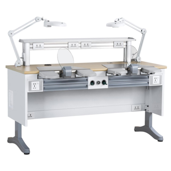
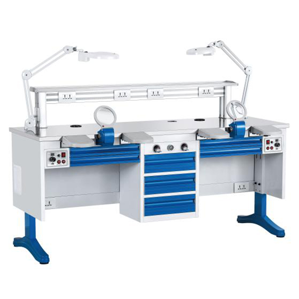
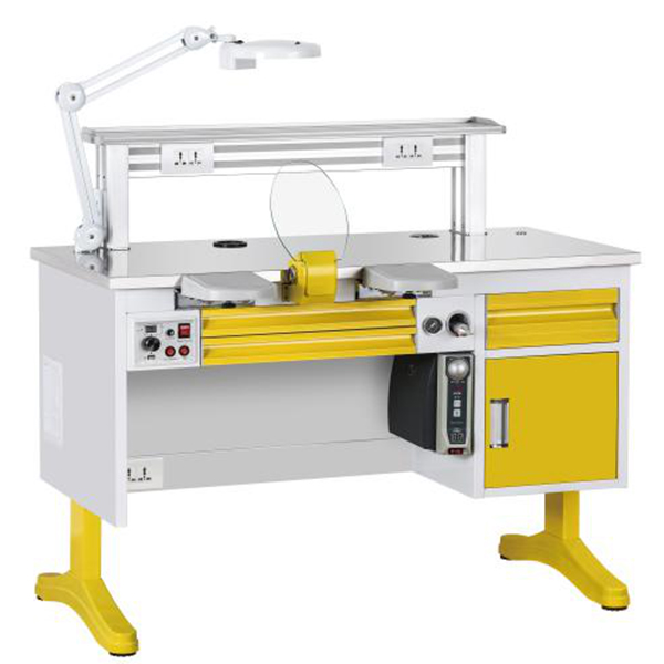
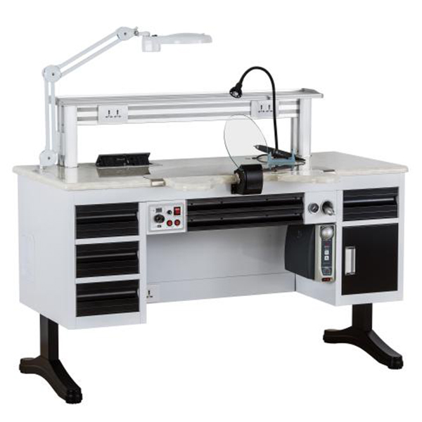
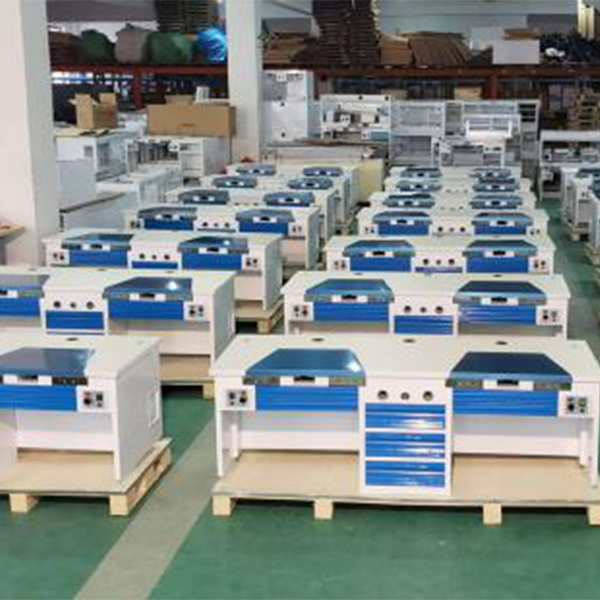
This type of Dental workstation widely adopts some famous dental technicians' advice both at home and abroad.The main material consists of cold-rolled stamping, die-casting aluminium alloy and extrusions.Through 3D modeling design, and by laser cutting machine,CNC bending machine, automatic welding machine and other modern equipment precision machining.Surface material can be made by squeezed high-density fireproof board, or stainless steel, or man-made marble.Selves on the desk are all made by aluminium alloy, which make the selves elegant and durable.The work light is combined by sun-light source and 5- inch 300-degree magnifier.Elbow guard surface is covered by elastomer material which makes the elbow more comfortable.The elbow working space can be adjusted in two levels between standard (40cm) and widen (48cm).Hand Gurad Layer Board adjustable any 5-20angle range.Self-reset air gun is well designed with good texture.The independent suction device also supports central vacuuming system which has strong power and low noise.The height of this desk can be adjusted between 80cm and 85cm.The four corners can be fine-tuned according to the ground.The delicate appearance air gun can reset automatic. The workstation configures low noise and suction power dust exhaust apparatus, also it support central dust collection system concentrated dust collection.Embedded integrated electrical control box is easy to fix and replace.This control box can control all of the electrical circuit, at the same time, based on this control box and from the digital window; workers can check the vacuum dust collection system of the motor run time.The standard configuration USB interface can support 2A 12V weak power.The suction nozzle assembly has two accessories can choose.One is Fan toughened glass block dust board another one is LED lamp ring (built-5 inch magnifying glass/5 inch flat lens). The workstation has a unique hidden layer, which is neat and easy to use, and can be used in the actual use of all pipelines, as well as vacuum cleaners are hidden in the sandwich.Between each workstations can connect seamlessly.There are supplementary dust collection components which are allocated randomly.The operation desk can be cleaned just by buckling the assistant suction device on the nozzle.
read more
OEM/ODM Supplier Dental Digital X Ray Sensor -
... OEM/ODM Supplier Dental Digital X Ray Sensor -
...
01

OEM/ODM Supplier Dental Digital X Ray Sensor - ...

2016-01-08
Pano: •Provide you with high diagnosis accuracy and better treatment on diseasesof the full dentiton and jaw. •Easy to get HD panoramic images by building a perfect tomographic track combined with the motion of the X, Y and Sita axis. CT : •A versatile helper to clearly display patient's anatomical structure with flexible FOV. •lntelligent imaging technology makes free observation from angle realizable. •Accurate measurement of the distance, area,volume and outline contour makes the one-key panoramic image obtained from the 3D image simple and easy. Cephalometric •Othodontic specialist's best choice making orthodontic treatment and maxillofacial surgery efficient and accurate. •Low-dose radiation and high gray realized by dual-level alignment adjustment to provide you with HD cephalometic images. •Unique algorithms of HRPBR image reconstruction and WE image processing •Leading technology intelligently generating HD pano images to provide with more clinical details and high diagnosis accuracy •Intelligent de-noise algorithm significantly improving image's resolution b yreducing image's noise and distortion •Variety of shooting modes: HD scanning, SD scanning, root canal scanning  •Special-designed algorithm of mental artifact removal providing with high quality images by removing artifacts caused by absorbent materials •CUDA high-speed image reconstruction High configuration and performance High Quality Flat Panel Detector Large surface and dynamic FPD ensure sensitivity and accuracy Metal Artifact Removal Effectively remove artifact caused by absorbent materials from the images with clearer details of human tissues displayed.
read more
Factory Cheap Hot Absorbent Paper Points -
 Api... Factory Cheap Hot Absorbent Paper Points -
 Api...
01

Factory Cheap Hot Absorbent Paper Points - Api...

2016-01-08
LASER FAST IMAGE PROCESSING Imaging is fast and simple. The smart top-loading design harnesses the power of gravity for simple loading and fuss-free use. In just a short time, the crystal clear radiograph appears on the computer screen, ready for viewing. Simply insert the next exposed plate when ready. FLEXIBLE IN MORE WAYS THAN ONE The Apixia DIGITAL PSP Scanner packs tremendous imaging power into a small package. It is tiny enough to fit into the most crowded dental operatories for handy chair-side use. Imaging with Phosphor plates offers all the flexibility and patient comfort of film, combined with the immediacy and environmental benefits of digital radiography. COMFORTABLE & REUSABLE PHOSPHER PLATES Imaging with Phospher Plates offers all the flexibility and patient comfort of film. Phospher plates are reusable a countless number of times to maximize your investment. 4 optional plate sizes allows for optimum size selection for each patient. SMART AND SIMPLE TOP-LOADING DESIGN Special top-loading design allows for easy image scanning. Just insert and scan any size plate for any type of clinical situation. Quick and easy scanning for easy image viewing on a computer display. 1.Small Tabletop Design 2.High Resolution Scans in a Fraction of the Time 3.Automatic In-line Eraser 4.Powerful, Easy-To-Use Software Included 5.Value-Priced for any Dental Practice Minimum Requirement for Computer Configuration: Windows 7/Windows 8/VISTA/XP (32, 64Bit) 1GB RAM Pentium 4 CPU or much advanced USB 2.0 transmit CD/DVD drive Resolution: >1024*768 40GB for hard drive space High resolution image display HD LCD This machine does not include the computer Digirex Psp Imaging Scanner Specification: Voltage: AC 100-240V, 50/60Hz Scanning time: 10s Resolution: 17 lp/mm Transfer way: USB 2.0 Size of digital X-ray plate: 0# 2# (1# 3# is optional) Built-in cleaning device: Automatic Size: 258*138*183mm Weight: 3.6kg
read more
Wholesale Price Base Plate Wax -
 Digital 3D OP... Wholesale Price Base Plate Wax -
 Digital 3D OP...
01

Wholesale Price Base Plate Wax - Digital 3D OP...

2016-01-08
Pano: •Provide you with high diagnosis accuracy and better treatment on diseasesof the full dentiton and jaw. •Easy to get HD panoramic images by building a perfect tomographic track combined with the motion of the X, Y and Sita axis. CT : •A versatile helper to clearly display patient's anatomical structure with flexible FOV. •lntelligent imaging technology makes free observation from angle realizable. •Accurate measurement of the distance, area,volume and outline contour makes the one-key panoramic image obtained from the 3D image simple and easy. Cephalometric •Othodontic specialist's best choice making orthodontic treatment and maxillofacial surgery efficient and accurate. •Low-dose radiation and high gray realized by dual-level alignment adjustment to provide you with HD cephalometic images. •Unique algorithms of HRPBR image reconstruction and WE image processing •Leading technology intelligently generating HD pano images to provide with more clinical details and high diagnosis accuracy •Intelligent de-noise algorithm significantly improving image's resolution b yreducing image's noise and distortion •Variety of shooting modes: HD scanning, SD scanning, root canal scanning  •Special-designed algorithm of mental artifact removal providing with high quality images by removing artifacts caused by absorbent materials •CUDA high-speed image reconstruction High configuration and performance High Quality Flat Panel Detector Large surface and dynamic FPD ensure sensitivity and accuracy Metal Artifact Removal Effectively remove artifact caused by absorbent materials from the images with clearer details of human tissues displayed.
read more